Education
The SEM Learning Center
Sub-epidermal moisture (SEM) technology has created a profound shift in modern pressure injury prevention. Learn why.
Get Insights in Minutes, Not Days
Watch How to Scan a Heel
Watch How to Scan a Sacrum
The How & Why of Pressure Injuries
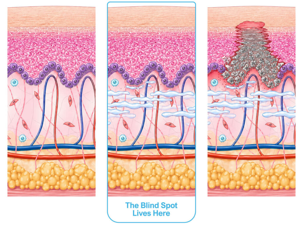
Pressure injuries begin with hidden inflammation and fluid buildup beneath the skin. With SEM scanning technology, clinicians have a clear window into early damage 5 days (median) before visible changes.1
Visual skin assessment has long been the foundation of pressure injury (PI) detection. Clinicians rely on it daily, and do so with genuine commitment to patient safety. But commitment cannot overcome what the method itself cannot provide: objective, biology-based detection of tissue damage before it becomes visible.
Subjective Results
Inconsistent Scoring
Limited Sensitivity
Dark Skin Tone Disparities
Not Localized
Hard to Measure & Defend
Subjective Results
Visual inspection is subjective and surface-level1. It reflects damage already in progress, not the earlier biological response that occurs beneath intact skin. Inflammation and fluid shifts can be underway long before there is anything to see.
Inconsistent Scoring
Reviews of the literature show clinician agreement on pressure injury staging is often poor to moderate, with Kappa values commonly in the range of about 0.3 to 0.6 among trained clinicians. Most studies report Kappa values clustering around 0.42. This means clinicians can reasonably disagree even when using the same definitions and having the same training. This variability reflects a method that depends on interpretation rather than measurement.
Limited Sensitivity
Systematic reviews of literature consistently show that visual skin assessment has low and variable sensitivity for detecting early tissue damage3. That's because pressure injury pathophysiology begins beneath intact skin before visible signs appear.
Dark Skin Tone Disparities
We are trying to look for redness, blanching or color changes. Skin redness can be difficult to identify in patients with darker skin tones4. This is a well observed and documented limitation of inherited systems. Early tissue damage can begin 3-10 days before it appears on the surface. When skin tone makes those changes harder to see, the first sign we recognize might already be stage 3 or stage 4 injury, and the data shows patients with darker skin tones are four times more likely to die from pressure injury related complications5.
Not Localized
Traditional prevention is largely built on proxies, and risk assessment tools estimate likelihood across the whole patient, not what is actually happening in the tissue at the specific anatomical sites most at risk, such as the sacrum and heels, which account for the majority of all pressure injuries.
Hard to Measure & Defend
When assessment depends on interpretation rather than objective measurement, variability is expected, and that variability contributes directly to delayed recognition, delayed intervention, and missed opportunities for prevention.
Pressure Injury Prevention Starts with Biology
Pressure injuries are a biological process that require biological detection. Prevention requires early detection, and early detection requires SEM scanning.
All Education Resources
Topic
Care Setting
SEM Scanning and eCQM HH-PI Documentation
Comprehensive Care for Joint Replacement eXpanded (CJR-X) Explained
Transforming Episode Accountability Model (TEAM) Explained
eCQM Hospital Harm – Pressure Injury Explained
SEM Delta™ (∆)
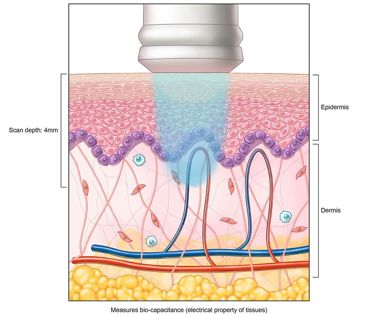
Introduction to Biocapacitance
SEM Assessment: Detection, Treatment and Prevention
Pressure Injury Classifications
Do No Harm
Explore proven solutions to achieve zero preventable harm.